


Magnetic Resonance Imaging (MRI): A Comprehensive Guide
Magnetic Resonance Imaging MRI
Understanding the intricacies of medical diagnostics can be daunting, but MRI technology has revolutionized the field, providing unparalleled insights into the human body. MRI scans have become a crucial diagnostic tool, enabling healthcare professionals to visualize internal structures with remarkable clarity. This non-invasive technique has transformed the diagnosis and treatment of various medical conditions.Key Takeaways
- Understanding MRI technology and its applications
- The significance of MRI in medical diagnostics
- How MRI scans aid in disease diagnosis and treatment
- The benefits of MRI over other imaging techniques
- What to expect during an MRI procedure
The Science and Evolution of MRI
Understanding MRI requires delving into its foundational principles and the technological advancements that have shaped it. Magnetic Resonance Imaging (MRI) is a sophisticated medical imaging technique that has revolutionized diagnostics.The Basic Principles of MRI
MRI operates on the principle of nuclear magnetic resonance. It uses a powerful magnetic field and radio waves to generate images of the body’s internal structures. When a patient undergoes an MRI procedure, they are placed within the MRI machine, which contains a strong magnetic field. This field aligns the hydrogen nuclei in the body, and radio waves are then used to disturb these alignments, creating signals that are picked up by the machine. These signals are then processed to create detailed images.Historical Development of MRI Technology
The development of MRI technology has been a gradual process, marked by significant milestones. The first MRI scan was performed in 1977, and since then, there have been numerous advancements, including improvements in magnetic field strength, radio wave technology, and image processing algorithms. These advancements have led to faster scan times, higher resolution images, and the ability to perform more complex scans. Today, MRI is a crucial diagnostic tool in medical practice, offering insights that were previously unimaginable.How Magnetic Resonance Imaging (MRI) Works
Magnetic Resonance Imaging (MRI) has revolutionized medical diagnostics by providing high-resolution images of the body’s internal structures without the use of ionizing radiation. The benefits of MRI include its ability to diagnose a wide range of conditions, from injuries and tumors to neurological disorders. Various MRI services are available, including functional MRI (fMRI) for brain activity assessment and MRI angiography for vascular imaging. Understanding the different types of MRI scans and their applications can help patients prepare for their procedures and know what to expect. The cost of MRI procedures can vary based on factors such as location, facility, and insurance coverage. It’s essential for patients to check with their healthcare providers and insurance companies to understand the costs involved. Interpreting MRI results requires a healthcare professional’s expertise. The results can provide critical information for diagnosing conditions, planning treatments, and monitoring disease progression. By understanding MRI benefits, services, cost, and results, patients can make informed decisions about their healthcare. Magnetic Resonance Imaging (MRI) plays a pivotal role in neurosurgery by providing detailed, high-resolution images of the brain and spinal cord, enabling precise diagnosis, surgical planning, and intraoperative guidance. Unlike other imaging modalities, MRI offers exceptional contrast between soft tissues, making it invaluable for identifying tumors, vascular malformations, traumatic injuries, and degenerative diseases affecting the central nervous system. Functional MRI (fMRI) and diffusion tensor imaging (DTI) further enhance surgical accuracy by mapping critical brain functions and white matter tracts, helping neurosurgeons avoid eloquent areas responsible for speech, movement, and cognition. Intraoperative MRI systems allow real-time imaging during surgery, reducing the risk of residual tumor and improving outcomes. Overall, MRI has revolutionized neurosurgery by enhancing safety, precision, and the ability to tailor interventions to individual patients’ neuroanatomy and pathology. Functional MRI (fMRI), which measures brain activity by detecting changes in blood oxygenation; Diffusion Weighted Imaging (DWI) and Diffusion Tensor Imaging (DTI), which assess the movement of water molecules and are crucial in stroke assessment and white matter tractography; Magnetic Resonance Angiography (MRA) for visualizing blood vessels; and Magnetic Resonance Spectroscopy (MRS) for evaluating metabolic changes in tissues. Additionally, Intraoperative MRI (iMRI) systems provide real-time imaging during neurosurgical procedures, enhancing surgical precision and minimizing residual pathology. These diverse MRI modalities have significantly advanced diagnostic accuracy, surgical planning, and treatment monitoring across a wide range of medical specialties.FAQ
What is Magnetic Resonance Imaging (MRI)?
Magnetic Resonance Imaging (MRI) is a non-invasive medical imaging technique that uses strong magnetic fields and radio waves to produce detailed images of the internal structures of the body.
How does an MRI machine work?
An MRI machine works by aligning the hydrogen atoms in the body using a strong magnetic field, and then disturbing them with radio waves to produce signals that are used to create detailed images.
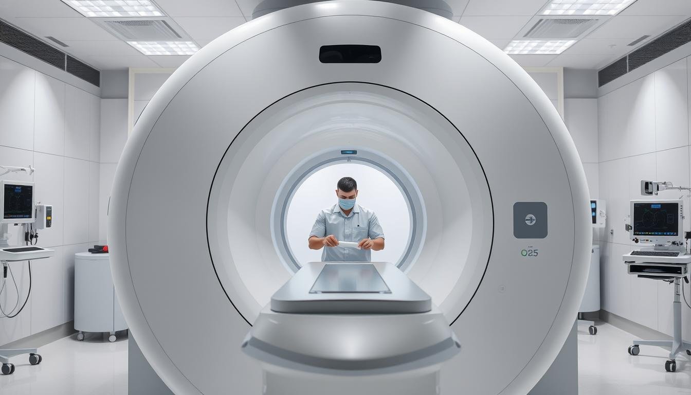
What is the MRI procedure like?
The MRI procedure typically involves lying on a table that slides into the MRI machine, which is a large, cylindrical tube. The procedure can take anywhere from 15 to 90 minutes, depending on the type of scan being performed.
What are the benefits of using MRI in medical diagnostics?
MRI provides high-resolution images without the use of ionizing radiation, making it a valuable diagnostic tool for a wide range of medical conditions, including injuries, infections, and tumors.
How much does an MRI scan cost?
The cost of an MRI scan can vary depending on factors such as the location, type of scan, and insurance coverage. On average, the cost of an MRI scan can range from 0 to ,000 or more.
How long does it take to get MRI results?
The time it takes to get MRI results can vary depending on the facility and the complexity of the scan. Typically, results are available within a few hours to a few days.
What are the different types of MRI scans?
There are several types of MRI scans, including functional MRI (fMRI), magnetic resonance angiography (MRA), and magnetic resonance venography (MRV), each with its own specific applications and benefits.